
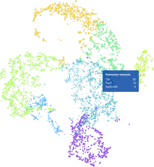
Single Gene Expression
Chromium Single Cell Gene Expression with Feature Barcode technology provides expression profiling of genes and proteins to genome editing, identify and characterize rare cell types and biomarkers, or examine therapeutic mechanisms of action a cell-by-cell basis with transcriptional profilingalone or multiomic characterization.
- Capture the full heterogeneity of a sample, in tens of thousands of cells.
- Measure 3’ mRNA, cell surface proteins, CRISPR perturbations, and more.
- Perfect to help exploring cellular heterogeneity, novel targets, and biomarkers with combined gene expression, surface protein expression, or CRISPR edits in each cell.
- Leverage sample multiplexing to maximize cell and sample throughput across species and sample types, such as fixed, fresh, and frozen samples, as well as whole cells and nuclei.
- Streamlined data analysis with one-day lab workflow

Single-Cell Immune Profiling
Chromium Single Cell Immune Profiling provides a comprehensive approach to simultaneously examine cellular heterogeneity of the immune system, T- and B-cell repertoire diversity, and antigen specificity at single cell resolution.
- Highly sensitive detection of 5’ mRNA with antigen specificity and phenotyping.
- Immune mapping by combining immune repertoire with antigen specificity at single cell resolution.
- Streamlined data analysis with one-day lab workflow
- Demonstrated on PBMCs, fresh and frozen cell samples, and dissociated tissue.

Single-Cell Multiome ATAC + Gene Expression
Chromium Single Cell Multiome ATAC + Gene Expression provides a unified view of a cell’s gene expression profile and its epigenomic landscape to increase the resolution of cell states, identify drivers of differential gene expression, and discover cells with similar transcriptional profiles but functionally different chromatin landscapes by leveraging two modalities at once.
- Profile gene expression and open chromatin from the same cell, across thousands of cells
- Understand drivers of tumor heterogeneity, mechanisms of therapeutic resistance, and the cell types that underlie neurodegenerative or immunological disorders.
- Discover new gene regulatory interactions and rare cell populations
- Streamlined data analysis and efficient lab workflow

Single-Cell ATAC
Chromium Single Cell ATAC (Assay for Transposase Accessible Chromatin) allows you to analyze chromatin accessibility at the single cell level, providing insights into cell types and states, and deeper understanding of gene regulatory mechanisms.
- Analyze thousands of unique open chromatin fragments per cell, genome-wide.
- Go beyond population averages by measuring epigenomic profiles in single nuclei.
- Profile hundreds to tens of thousands of nuclei per chip.
- Demonstrated with cell lines, primary cells, and fresh and frozen tissue samples.
- Streamlined data analysis with one-day lab workflow
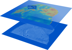
Spatial Gene Expression
Visium Spatial Gene Expression is a next-generation molecular technology for classifying tissue based on total mRNA. Map the whole transcriptome with morphological context to discover novel insights into normal development, disease pathology, and clinical translational research.
- Combine whole transcriptome spatial analysis with immunofluorescence protein detection.
- Analyze the whole transcriptome from an entire section.
- Offer high resolution depending on tissue type (1–10 cell resolution on average per spot)
- Demonstrated on a diverse set of organs across species (human, mouse, rat, and more).
- Streamlined data analysis with one-day lab workflow
*Now for FFPE tissue
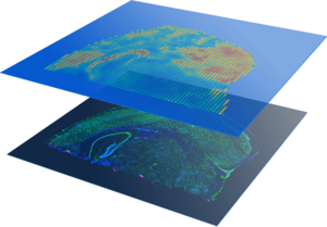
Spatial Proteomics
Spatial Gene Expression with Immunofluorescence can visualize spatial patterns of gene expression together with protein detection by immunofluorescence on the same tissue section. Histological tools like in situ hybridization and immunofluorescence alone are limited in the breadth of analysis they can perform. The Visium Spatial Gene Expression Solution with immunofluorescence enhances these techniques, letting researchers to combine immunofluorescence protein detection and unbiased, spatial gene expression in the same tissue section alongside histological analysis.
- Ready-to-use, robust workflow for whole tissue section analysis
- Easy to integrate with current histological laboratory methods and tools for tissue analysis
- Antibody flexibility: Simultaneously visualize protein and RNA using your own validated antibodies
- Broaden the reach beyond predefined regions of interest to analyze the entire tissue section at once
- Detect more analytes with whole transcriptome plus up to 6 proteins simultaneously